Assess
Start with the clinical information and visual data available at that moment.
TraumaVault contains authentic trauma teaching material and clinical imagery. Some cases may include severe injuries, blood, open wounds, exposed tissue, operative findings or other graphic content that may be disturbing.
By entering, you confirm that you are at least 18 years old, are accessing this material for professional medical education, and understand that you may encounter graphic trauma imagery.
TraumaVault is built around the moment that matters most in trauma and acute care surgery: the decision. Study an image, read the evolving case, commit to an action, then see how the case unfolds.
Start with the clinical information and visual data available at that moment.
Choose what you would actually do before the answer is revealed.
See the reasoning, consequences, and next stage of the evolving case.
Track your decisions and earn up to 100 points for each case.
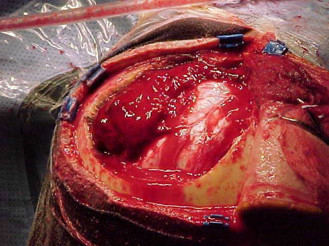
CASE 001A 45-year-old woman is brought to the emergency department after an unwitnessed fall down a flight of stairs while intoxicated. She has a GCS of 5, unequal pupils with a sluggishly reactive left pupil, and blood in the external auditory canal. She is intubated, cervical-spine precautions are maintained, and initial resuscitation is performed. After stabilization sufficient for imaging, head CT demonstrates a large acute epidural hematoma with marked mass effect, midline shift, and concern for impending herniation. Neurosurgery is contacted, and the patient is transferred emergently to the operating room. The operative image becomes available after cranial exposure and evacuation have begun.
 CASE 002
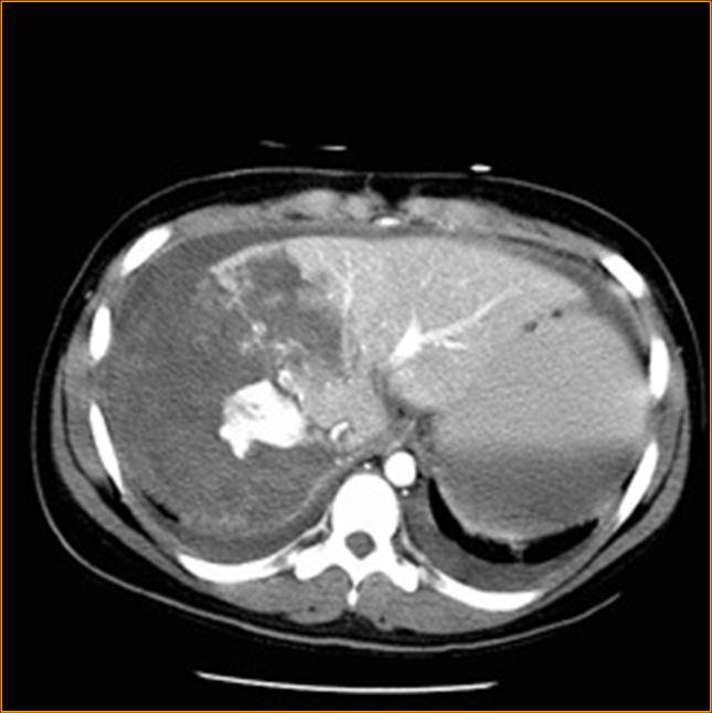
A 29-year-old restrained driver is evaluated after a high-speed MVC with right-upper-quadrant pain. After initial resuscitation he is sufficiently stable for contrast-enhanced CT of the abdomen and pelvis.
 CASE 003
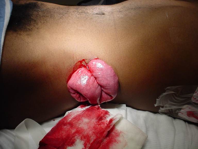
A 29-year-old patient is brought directly to the trauma bay shortly after being stabbed in the left flank during an assault. The patient is awake, speaking clearly, and protecting the airway. Initial vital signs are heart rate 108/min, blood pressure 118/74 mmHg, respiratory rate 20/min, and oxygen saturation 98% on room air. During the initial external examination, an approximately 4-cm left flank wound is identified with bowel protruding through the opening; the accompanying image was obtained at this examination and is available now. No active external arterial bleeding is seen. The abdomen is tender near the wound, but there is no diffuse rigidity documented on the initial examination.
 CASE 004
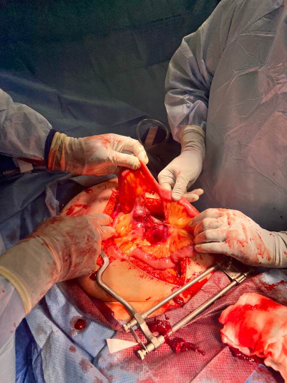
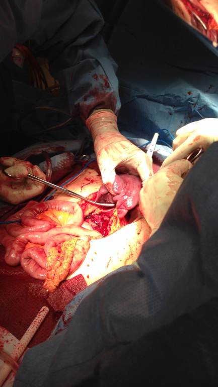
An adult restrained driver is brought after a high-speed motor-vehicle collision. There is a prominent lower abdominal seatbelt sign and abdominal tenderness. Initial airway and breathing assessments are adequate. Circulation is supported with blood products and limited crystalloid; the patient remains responsive for immediate trauma assessment but develops worsening abdominal pain and borderline perfusion. FAST demonstrates free intraperitoneal fluid. Because the patient has evolving perfusion concern and a positive FAST, the trauma team proceeds to emergency exploratory laparotomy rather than delaying for CT. The supplied operative photograph is obtained after the abdomen is opened.
 CASE 005
An adult patient is brought to the trauma bay after a high-speed motor-vehicle collision. On arrival, the patient is pale, confused, and hypotensive with abdominal tenderness and guarding. Initial resuscitation includes blood products, large-bore vascular access, and activation of the massive transfusion protocol. eFAST demonstrates free intraperitoneal fluid in the right upper quadrant and pelvis. The patient remains persistently hypotensive despite initial resuscitation, so the trauma team proceeds directly to emergent exploratory laparotomy without CT. The uploaded operative photograph becomes available after abdominal entry and exposure.
 CASE 006
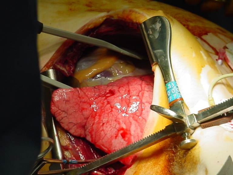
A previously responsive adult is brought after a high-speed motor-vehicle collision. Initial resuscitation includes airway control, bilateral chest decompression as indicated by the trauma team, blood-product resuscitation, and rapid assessment for immediately reversible causes. The patient then loses pulses in the trauma suite. Because the patient is in traumatic arrest, the team proceeds directly to an adjacent operating room or operating-level resuscitation area rather than CT, initiates traumatic-arrest protocols, and performs an emergency resuscitative thoracotomy. The pericardium is opened, and the operative image becomes available during the procedure. The team now needs coordinated decisions about internal cardiac massage, rhythm assessment, and use of internal shock paddles.
 CASE 007
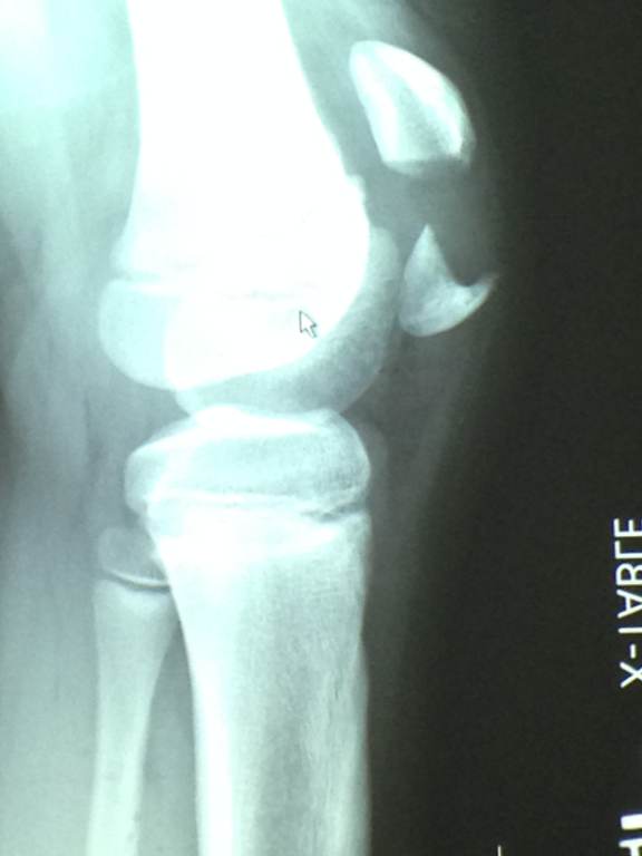
A 42-year-old adult presents to the emergency department after falling directly onto the front of the right knee. The patient is hemodynamically stable and has isolated knee pain and swelling. The skin is intact, distal pulses and sensation are normal, and there is no evidence of compartment syndrome. No knee radiographs have yet been reviewed in the case.
 CASE 008
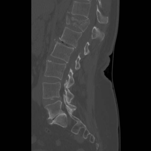
A 34-year-old restrained driver is brought to the emergency department after a frontal motor vehicle collision. The patient has a prominent transverse abdominal ecchymosis consistent with a seatbelt sign, severe low-back pain, and diffuse abdominal tenderness without peritonitis. Initial airway and circulation are intact. After analgesia and initial resuscitation, vital signs remain adequate for CT. Trauma CT imaging is obtained, including a sagittal lumbar reconstruction. The uploaded CT image is now available for review.
 CASE 009
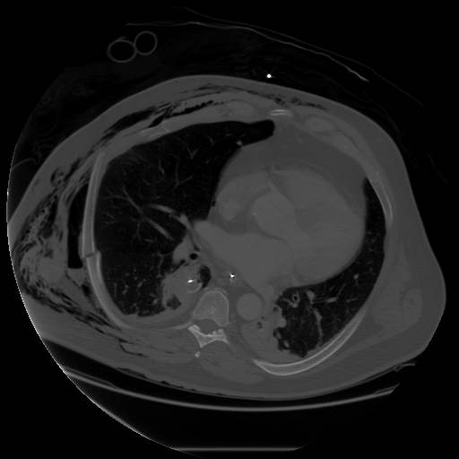
A restrained adult is brought after a high-speed MVC with a crushed chest. On arrival, there is marked chest-wall crepitus and subcutaneous emphysema, and the patient is intubated in the trauma bay. The patient is tachycardic but initially maintains blood pressure after blood products and controlled ventilation are initiated. After immediate trauma-bay reassessment and pleural decompression as clinically indicated, a right thoracostomy tube is placed. Oxygenation and blood pressure remain temporarily adequate, so the patient undergoes contrast-enhanced CT of the chest as part of the trauma CT evaluation. The uploaded CT image is now available for interpretation.
 CASE 010
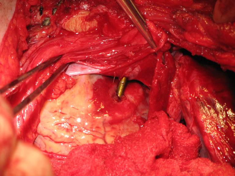
A worker is struck in the precordium by a nail fired from a pneumatic nail gun. The nail remains embedded and is not manipulated in the field. On arrival, he is conscious but anxious, with tachycardia, hypotension, muffled heart sounds, and a left parasternal wound. He has a weak but present pulse and transient improvement after blood products. A focused cardiac ultrasound shows pericardial fluid and a linear echogenic foreign body traversing the cardiac silhouette. Because of ongoing concern for cardiac penetration and marginal but temporarily supported hemodynamics, the trauma team proceeds urgently to the operating room with the nail stabilized in place. After thoracotomy and exposure of the heart, the operative image becomes available.
 CASE 011
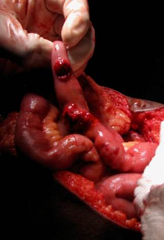
A patient arrives after a gunshot wound to the abdomen with hypotension and clinical concern for intra-abdominal hemorrhage and contamination. After immediate airway and circulation support with blood-product resuscitation, the trauma team proceeds directly to the operating room for emergent exploratory laparotomy. The uploaded image becomes available after the abdomen is opened and the small bowel is examined, showing multiple perforations with gross enteric leakage.
 CASE 012
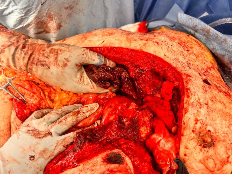
A 29-year-old restrained driver is brought after a high-speed motor-vehicle collision. On arrival, the patient is pale, confused, and hypotensive with abdominal distention and diffuse tenderness. Initial blood-product resuscitation produces only transient improvement, but the patient remains sufficiently responsive to proceed directly to the operating room. ED FAST is positive in the right upper quadrant and pelvis. The trauma team performs an emergency exploratory laparotomy. At operative exposure, the uploaded image becomes available: the abdomen contains massive hemoperitoneum, and the liver is visibly split into two major portions with active diffuse hemorrhage. The immediate operative challenge is temporary hemorrhage control rather than definitive hepatic reconstruction.
 CASE 013
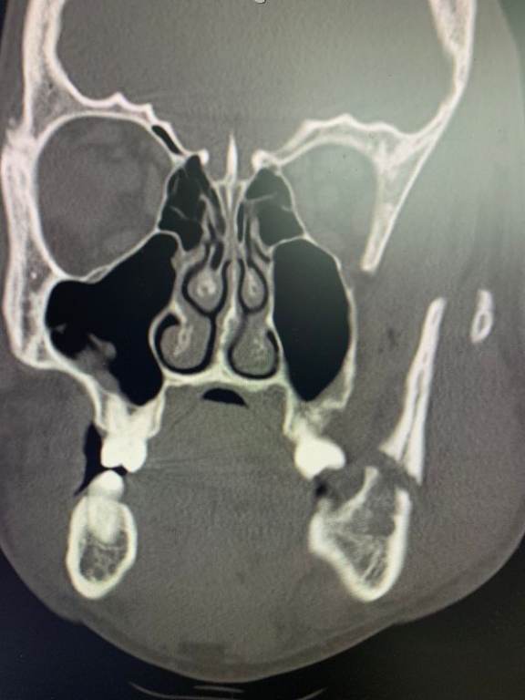
A 29-year-old man is brought to the emergency department after being repeatedly punched in the face during a bar fight. He is alert, speaking clearly, and breathing without difficulty. Initial assessment shows no stridor, expanding neck hematoma, or uncontrolled oral bleeding. He has facial swelling, malocclusion, pain with jaw movement, and a small intraoral laceration. Analgesia is provided, and cervical-spine precautions are maintained until examination is reassuring. Because he remains hemodynamically stable and has focal mandibular tenderness with malocclusion, diagnostic CT of the maxillofacial bones is obtained. The uploaded coronal CT image is now available for review.
 CASE 014
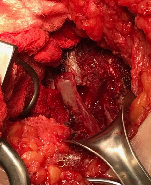
An adult patient is brought to the trauma center after a high-energy motor-vehicle collision with a crushed chest. Initial assessment shows respiratory failure, paradoxical chest-wall motion, severe chest pain before sedation, and hypoxemia. The patient is intubated, mechanically ventilated, and admitted to the trauma ICU. After initial resuscitation, CT chest demonstrates multiple markedly displaced fractures involving several contiguous ribs with a flail segment and pulmonary contusions; no injury requiring emergent abdominal surgery is identified. The patient remains hemodynamically suitable for further evaluation and planned intervention. Despite lung-protective ventilation, analgesia, pulmonary toilet, and supportive care, chest-wall instability and ventilator dependence persist. On the following hospital day, the patient is taken to the operating room for planned rib stabilization. After induction, positioning, and open exposure of the injured chest wall, the teaching image is obtained before fixation is completed.
 CASE 015
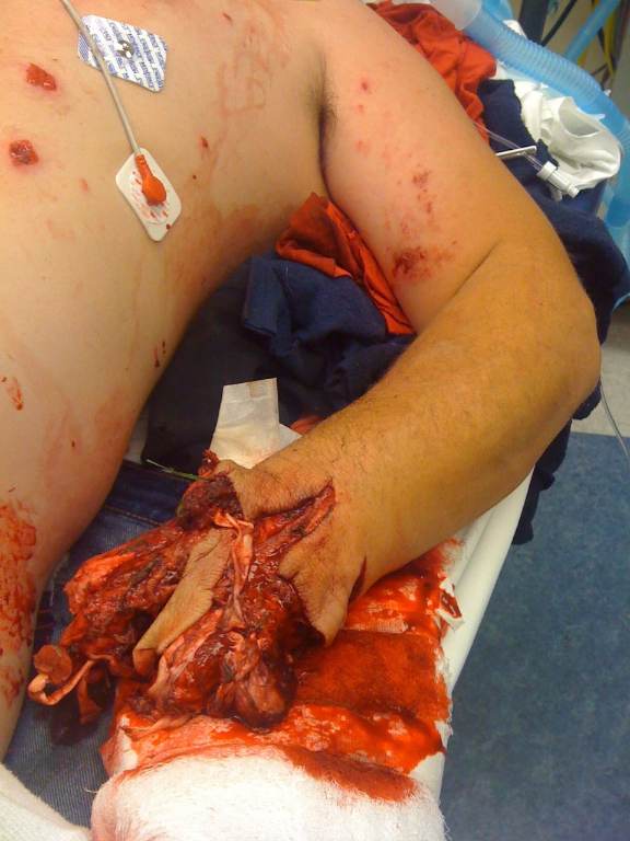
A previously healthy adult presents immediately after an M80 detonated in his hand. He is awake but distressed. The injured hand has near-complete soft-tissue disruption, gross contamination, and ongoing brisk bleeding through field dressings. On arrival, the trauma team obtains the supplied external photograph during the initial resuscitation; no CT or operative procedure has occurred. The team is evaluating airway, breathing, circulation, associated injuries, and immediate limb-threatening hemorrhage while arranging urgent specialist involvement.
 CASE 016
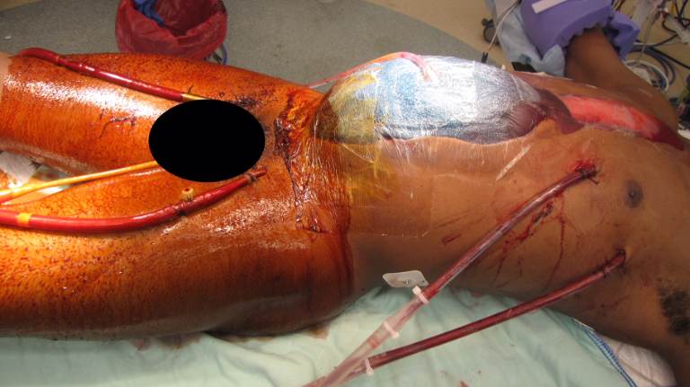
A previously healthy adult sustains a close-range gunshot wound to the right lower chest and upper abdomen. Prehospital care includes hemorrhage control, intubation, blood products, and rapid transport. In the trauma bay, the patient has penetrating thoracoabdominal injuries with hemorrhagic shock. Resuscitation proceeds directly to emergent operative management because of instability. Damage-control thoracotomy and laparotomy identify a transdiaphragmatic ballistic tract involving the lung and liver with major hemorrhage. The abdomen is packed and left temporarily open. Despite operative hemorrhage control and massive transfusion, severe hypoxemia and cardiopulmonary shock persist; the patient is cannulated for ECMO in the operating room and transferred to the ICU. The teaching photograph is obtained after this operative pathway, either during the operation or in the early postoperative ICU period, and becomes available at the second decision point.
 CASE 017
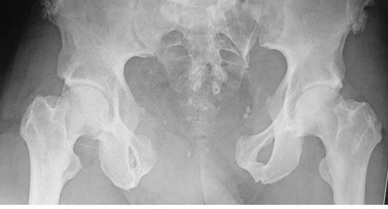
A 34-year-old restrained driver is brought directly to the trauma bay after a high-speed MVC. He is pale, confused, and complains of severe pelvic pain. Initial assessment shows a patent airway, spontaneous breathing, diminished but present distal pulses, and no obvious external hemorrhage. A pelvic binder has not yet been applied. After direct-pressure dressings, warming, large-bore access, and initiation of balanced blood-product resuscitation, an AP pelvic radiograph is obtained in the resuscitation bay because pelvic hemorrhage is suspected. The patient remains hypotensive but has transient improvement in mental status and radial pulse after the first blood products, permitting image acquisition. The radiograph is now available for interpretation.
 CASE 018
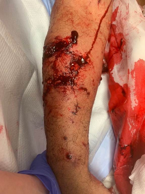
A 34-year-old right-hand-dominant adult presents to the emergency department approximately 2 hours after being attacked by an unfamiliar dog. Bleeding was controlled with a pressure dressing before arrival. The dog cannot immediately be located for verification of vaccination status. On arrival, the patient is alert and hemodynamically stable. The right arm has several deep punctures and lacerations with gross contamination and swelling. During the initial examination, the clinician obtains the displayed external wound photograph; it is not an operative image or diagnostic scan. Before analgesia, the patient reports numbness along the dorsoradial forearm and difficulty extending the wrist and fingers. Radial and ulnar pulses are palpable, capillary refill is normal, and the hand is warm.
 CASE 019
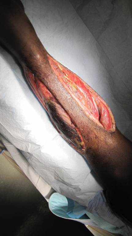
A 29-year-old construction worker sustains a heavy crush injury to the right lower leg when trapped briefly between machinery components. Plain radiographs at the receiving trauma center show a closed tibial shaft fracture. After splinting, analgesia, and initial resuscitation, serial examinations show escalating pain despite opioids, pain with passive toe stretch, tense compartments, and worsening paresthesia. Distal pulses remain palpable. The orthopedic trauma team is urgently consulted, and the patient proceeds to the operating room for suspected acute compartment syndrome. The uploaded photograph is obtained intraoperatively after the fasciotomy and becomes available during postoperative handoff; it is not used to establish the preoperative diagnosis.
 CASE 020
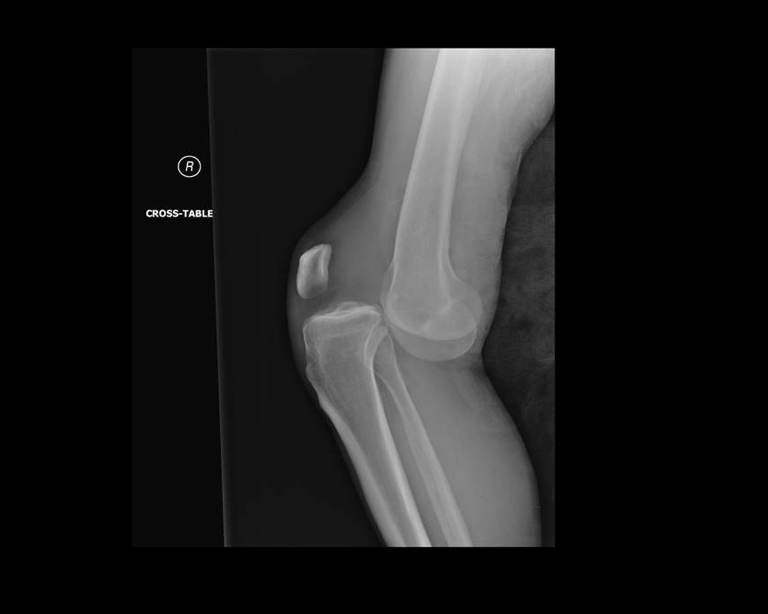
A 29-year-old restrained driver is brought to the emergency department after a high-speed frontal collision. The right knee struck the dashboard. The leg is held in flexion with a prominent anterior contour at the knee and severe pain. There is no uncontrolled external hemorrhage. After spinal precautions, analgesia, and initial trauma assessment, the patient is awake and physiologically stable. A portable cross-table lateral radiograph of the right knee is obtained in the ED before reduction and transferred to the trauma workstation; the uploaded image is revealed at Stage 1. The foot is warm, and a dorsalis pedis pulse is palpable but diminished compared with the left; the posterior tibial pulse is difficult to appreciate. Motor function is limited by pain, and sensation is reduced subjectively over the dorsum of the foot.
 CASE 021
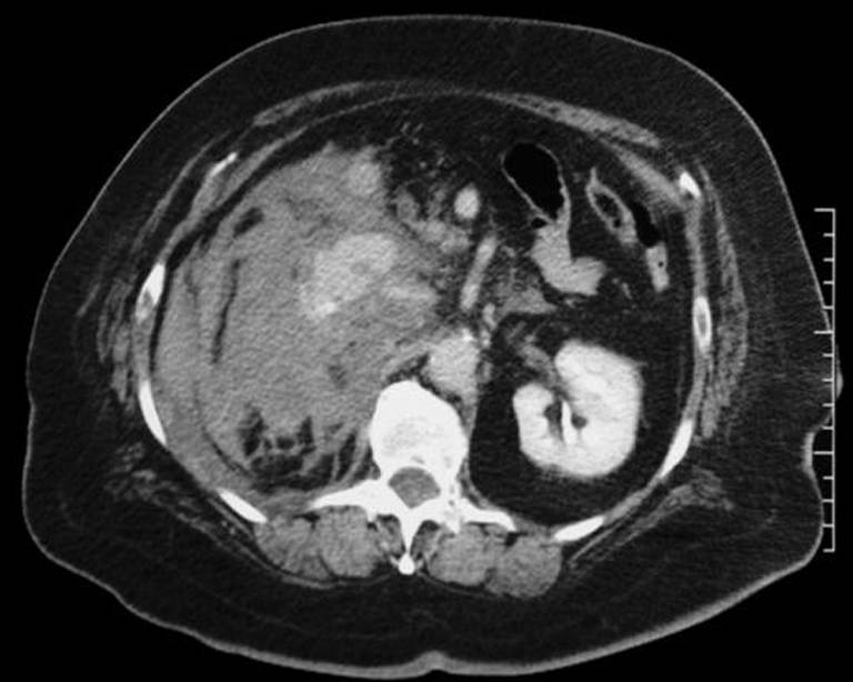
A motorcyclist is thrown from his bike at high speed. He arrives immobilized with right flank and abdominal pain, abrasions, and no obvious external hemorrhage. Initial airway and breathing assessments are reassuring. After warmed blood products and limited crystalloid, his systolic blood pressure is approximately 110 mmHg, heart rate 118/min, and mental status is normal. Pelvic stability is maintained, and there is no clinical evidence of tension pneumothorax. Because he remains sufficiently responsive to resuscitation, a contrast-enhanced trauma CT is obtained. The uploaded CT image becomes available for review after acquisition. Immediately after leaving the scanner, he becomes pale and confused, and his systolic blood pressure falls to 60 mmHg.
 CASE 022
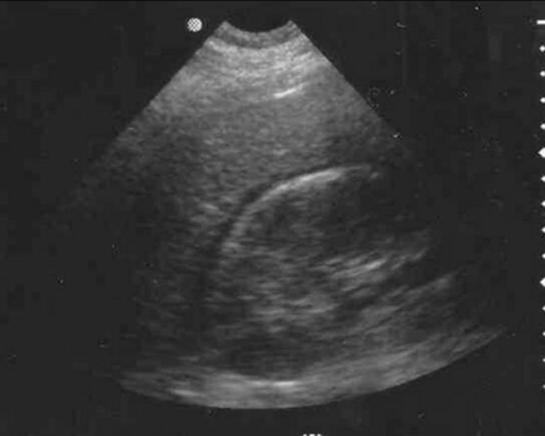
A 29-year-old patient arrives after a restrained high-speed motor-vehicle collision. The patient has right-upper-quadrant abdominal tenderness but no peritonitis. Initial airway and breathing assessments are normal. After a brief trauma-bay assessment, blood pressure remains 124/78 mmHg, heart rate 96/min, mental status is normal, and peripheral perfusion is adequate. A FAST examination is performed during the initial resuscitation; the supplied FAST image becomes available at the first decision point.
 CASE 023
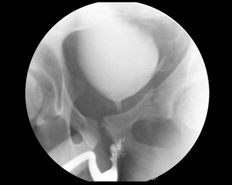
A 27-year-old man is brought after a motorcycle collision. He is alert, with pelvic pain and visible pelvic deformity. Initial blood pressure is 108/72 mmHg and heart rate is 116/min. After warmed blood products and pelvic stabilization, blood pressure improves to 124/80 mmHg. Examination shows blood at the urethral meatus, and the pelvis is not repeatedly stressed. Because urethral injury is suspected, a retrograde urethrogram is obtained before any urethral catheter attempt. The patient remains sufficiently stable for imaging, and the image is now available for interpretation.
 CASE 024
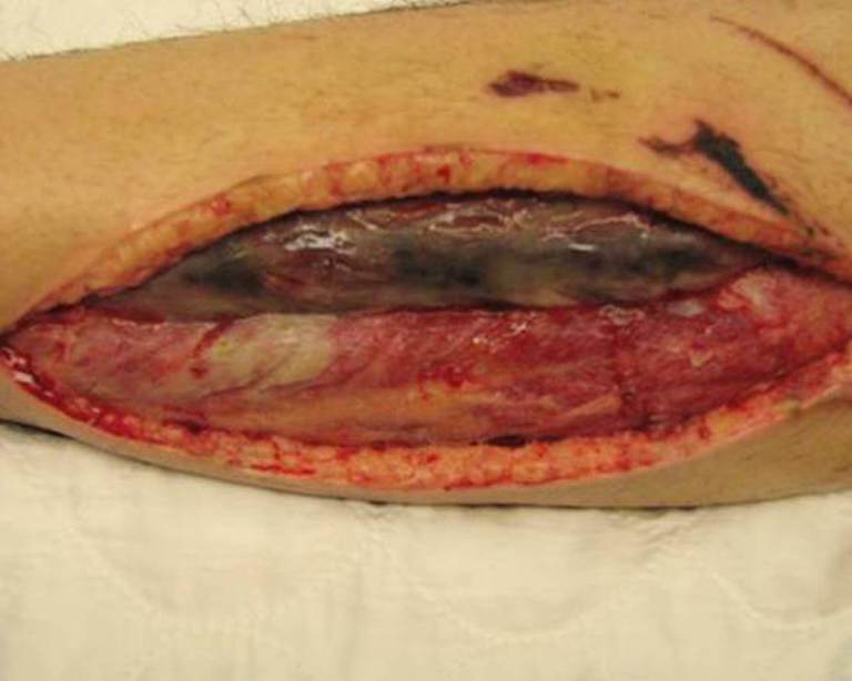
A 34-year-old patient sustains a high-energy crush injury to the lower leg. After examination confirms acute compartment syndrome, emergent four-compartment fasciotomy is performed. The wounds are left open, and the patient initially stabilizes. Forty-eight hours later, the patient develops fever to 39.1°C, worsening leukocytosis, increasing pain and malodor from the fasciotomy wound, and tissue discoloration. The patient remains sufficiently stable for immediate transfer to the operating room. The wound is taken for urgent repeat exploration, and the operative image becomes available after exploration begins.
 CASE 025
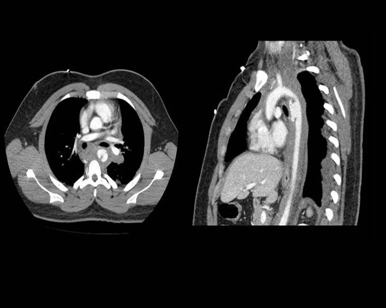
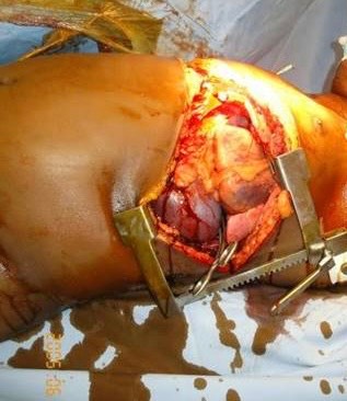
A 34-year-old restrained driver is brought to the emergency department after a high-speed frontal MVC. His chest struck the steering wheel. He is initially alert with chest and interscapular pain. Following the primary survey, initial hemorrhage-control measures, and analgesia, he has no refractory shock and remains sufficiently stable for diagnostic imaging: blood pressure 148/86 mmHg, heart rate 104/min, and oxygen saturation 97% on supplemental oxygen. There is a seatbelt mark and anterior chest tenderness without obvious external hemorrhage. A contrast-enhanced CT angiogram of the chest has been obtained after this initial assessment and is now available for review.
 CASE 026
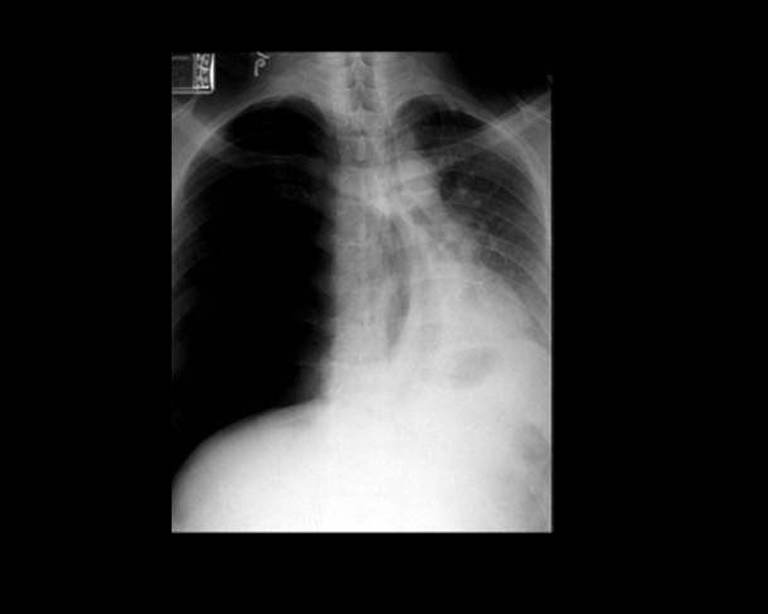
A 29-year-old restrained driver presents after a high-speed motor-vehicle collision. He is initially awake and speaking, with right-sided chest discomfort and mild dyspnea. Initial assessment shows a patent airway and no obvious external chest wound. After initial stabilization sufficient to obtain bedside radiography, a portable AP chest radiograph is obtained. The image is now available for review. The pre-radiograph examination documented reduced right-sided breath sounds, hyperresonance to percussion, increasing respiratory effort, and subtle tracheal deviation toward the left.
 CASE 027
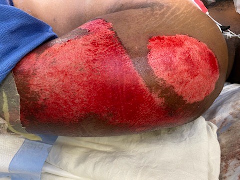
A 27-year-old motorcycle rider is brought to the emergency department after being thrown from the bike and sliding approximately 20 meters on asphalt. He was helmeted, awake, and hemodynamically stable throughout transport. After arrival, the primary survey shows no airway, breathing, or circulation compromise. During the initial ED exposure examination, before analgesia, irrigation, or debridement, a clinician obtains the uploaded photograph. It shows extensive road rash over the lateral hip and buttock. He also has scattered abrasions on the forearm and knee, but no obvious deformity. The wound is painful and visibly contaminated with fine particulate debris. The photograph does not exclude occult fracture, dislocation, neurovascular injury, compartment syndrome, or deeper soft-tissue injury.
 CASE 028
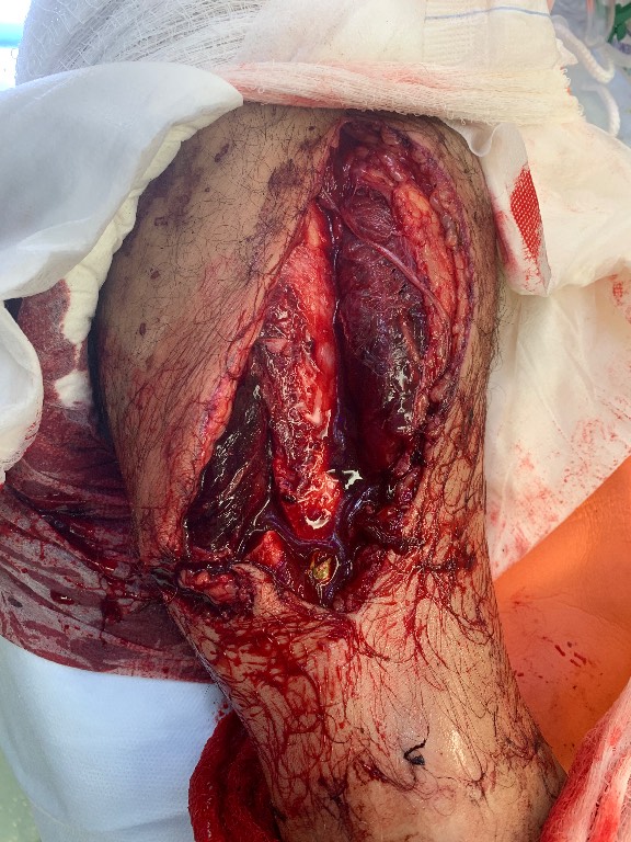
A 45-year-old motorcyclist is brought to the trauma bay after a high-speed collision. The left thigh has a grossly contaminated open injury with embedded road debris and obvious deformity. Distal pedal pulses are present but diminished compared with the opposite side; motor and sensory examination is limited by pain. There is no uncontrolled external arterial hemorrhage. After hemorrhage control, splinting, and initial blood-product resuscitation, the patient is sufficiently stable for urgent operating-room transfer. The wound is covered with sterile saline-moistened dressings, the limb is gently aligned and splinted, antibiotics are prepared for immediate administration, and tetanus status is assessed. The operative photograph is not available until the patient reaches the operating room.
 CASE 029
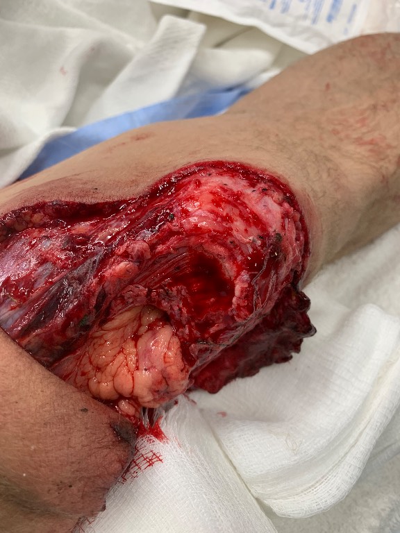
A 29-year-old patient is brought to the trauma bay after a high-speed MVC. The left knee has a large open wound with substantial tissue loss and contamination. Distal pulses are palpable, the foot is warm, and there is no uncontrolled external hemorrhage. Initial airway and breathing assessments are reassuring. After hemorrhage control with direct pressure, sterile dressing placement, analgesia, and IV access, the patient remains sufficiently stable for focused extremity evaluation and urgent orthopedic consultation. Once the wound is exposed for examination, the trauma team obtains the supplied external photograph. The image shows a deep, extensive open knee injury with exposed and devitalized tissue. The immediate priorities are infection prevention, protection of the wound, neurovascular surveillance, and operative evaluation.
 CASE 030
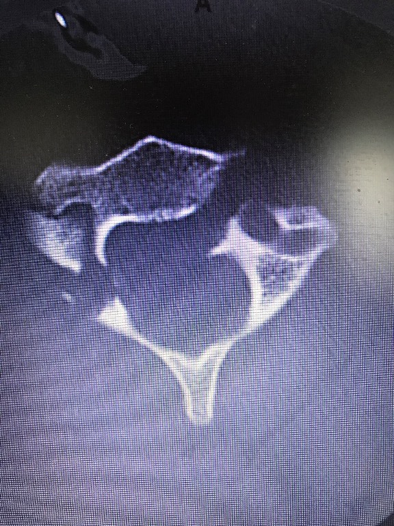
A 34-year-old restrained driver is brought to the emergency department after a high-speed side-impact collision. He was initially alert but reports severe neck pain and paresthesias in the left hand. He is hemodynamically stable, breathing spontaneously, and has no focal motor deficit. A cervical collar is maintained. After initial trauma assessment, CT of the cervical spine is obtained because of the high-risk mechanism and neurologic symptoms. The uploaded axial bone-window image is now available for review.
 CASE 031
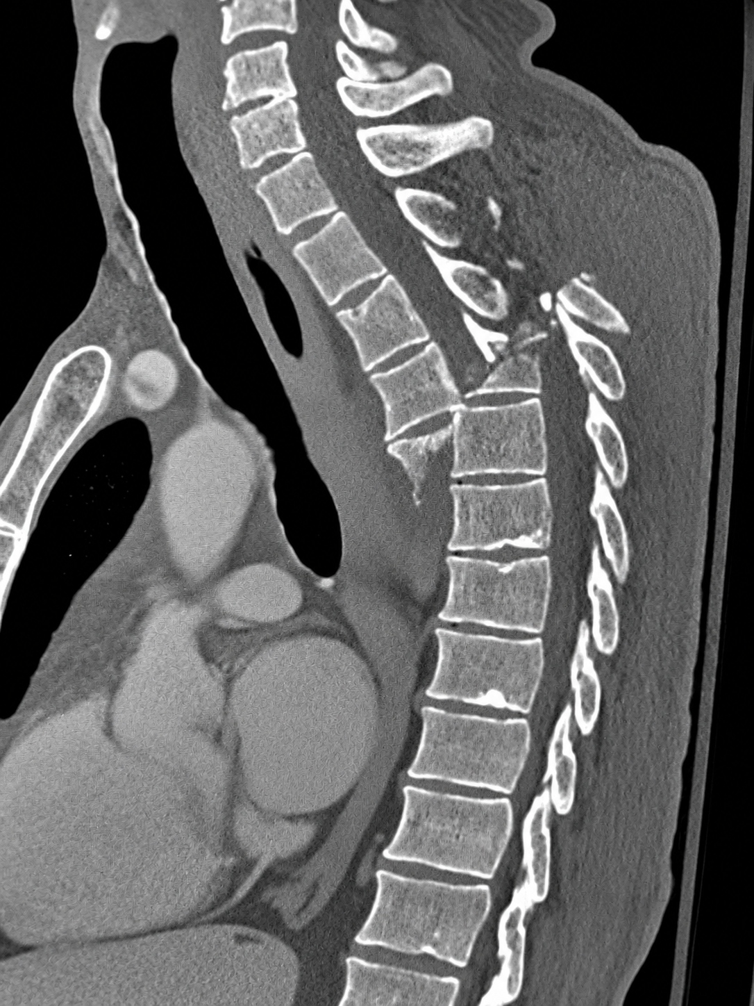
A 29-year-old patient is brought to the emergency department after a high-speed collision. The patient was extricated with manual in-line stabilization and remains immobilized. On arrival, the airway is patent, breathing is adequate, and peripheral pulses are present. After initial resuscitation, blood pressure is 108/70 mmHg, heart rate is 104/min, and oxygen saturation is 98% with supplemental oxygen. The patient is awake but frightened and reports severe neck and upper-back pain. The initial assessment does not identify an immediate indication for emergency operative intervention before diagnostic imaging. The patient is transferred with continuous monitoring and spinal precautions for CT.
 CASE 032
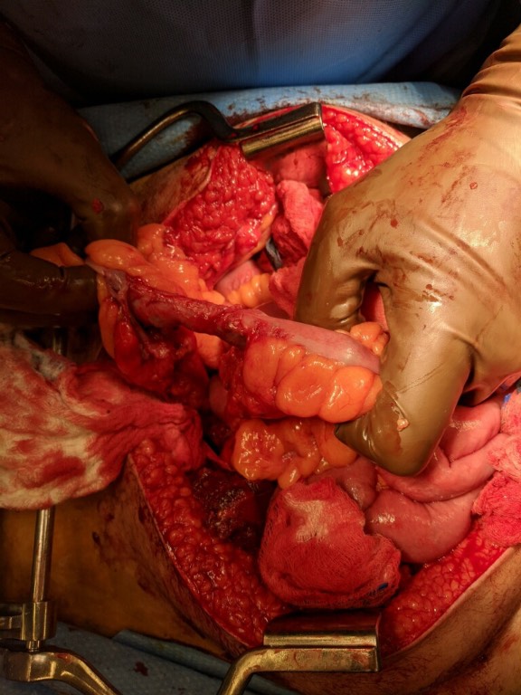
An adult involved in a high-speed motor-vehicle collision arrives with abdominal bruising in a seatbelt distribution. The patient is hypotensive and has altered mental status, so rapid-sequence intubation is performed in the resuscitation bay. Focused abdominal ultrasonography is positive for free intraperitoneal fluid. After a brief damage-control resuscitation with blood products, the patient remains unstable but can be transferred directly to the operating room for exploratory laparotomy. CT is not obtained because it would delay hemorrhage control. The operative image is acquired only after the abdomen is opened and the small bowel is examined.
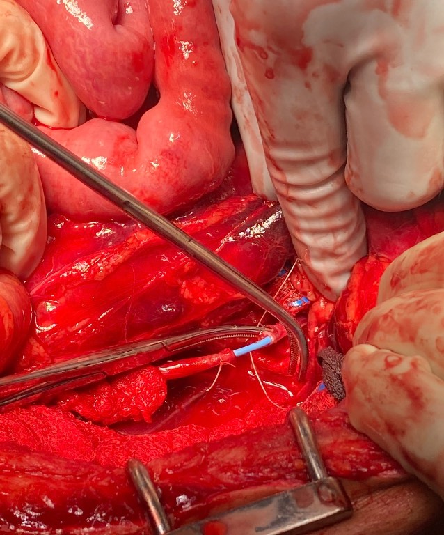 CASE 033
A 29-year-old man arrives after a close-range gunshot wound to the left flank. He is initially hypotensive but responds to blood products and remains sufficiently stable for focused assessment and contrast-enhanced CT. CT demonstrates a left retroperitoneal bullet tract with urinary extravasation and a perinephric hematoma; there is no clear major renal hilar disruption. He undergoes urgent exploratory laparotomy with retroperitoneal exploration. The operative photograph becomes available during repair of the identified ureteral injury.
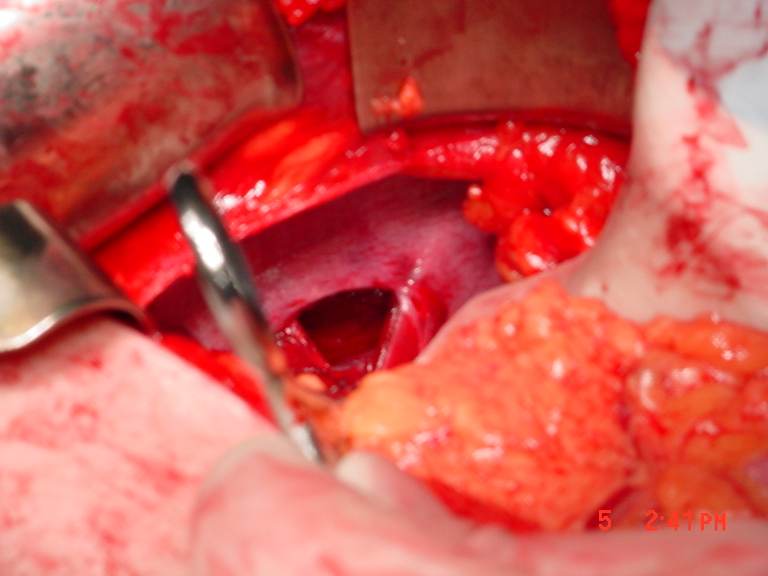 CASE 034
A 34-year-old pedestrian is run over by a vehicle. On arrival, the patient is awake but tachycardic, with left-sided chest pain, upper abdominal tenderness, and reduced breath sounds at the left base. Initial resuscitation with blood products and limited crystalloid improves perfusion sufficiently for diagnostic imaging. Chest and abdominal plain films suggest an abnormal left hemidiaphragm with possible herniation. Contrast-enhanced CT then demonstrates a traumatic left diaphragmatic defect with herniation of abdominal contents into the thorax, without an immediately lethal aortic injury. Because the patient remains adequately perfused and the defect requires definitive management, the trauma team proceeds to the operating room. After laparotomy, reduction of the herniated contents, and direct exposure of the diaphragm, the operative image becomes available.
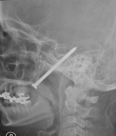 CASE 035
An adult patient is brought to the emergency department after an apparent self-inflicted nail-gun injury through the hard palate while intoxicated. She is awake, following commands, and protecting her airway, although speech is dysarthric from the oral injury. There is oral bleeding without uncontrolled external hemorrhage. Initial airway, breathing, and circulation assessment is stable, and cervical spine precautions are maintained. A lateral radiograph was obtained during the ED evaluation and is now available for review. It demonstrates a retained nail entering through the hard palate with a trajectory toward the skull base. The patient has no focal neurologic deficit on the initial examination.
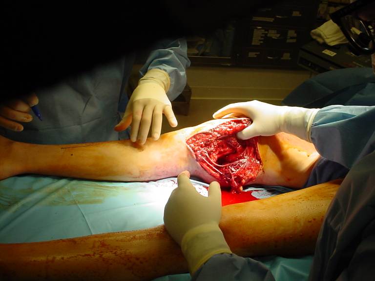 CASE 036
A 27-year-old patient arrives after a close-range gunshot wound to the posterior right knee. A prehospital tourniquet was applied approximately 1 hour before arrival for brisk bleeding. On resuscitation, the patient is awake but pale, with improving blood pressure after blood products. The tourniquet remains in place because bleeding recurs when it is loosened. The right foot is cool and pale with absent dorsalis pedis and posterior tibial Doppler signals. There is a penetrating wound in the popliteal fossa and expanding soft-tissue swelling. The patient has no other immediately life-threatening injuries identified on the initial trauma survey. After balanced blood-product resuscitation, the patient is sufficiently stabilized for urgent transfer to the operating room; CT angiography is not required before operative hemorrhage control and revascularization.
 CASE 037
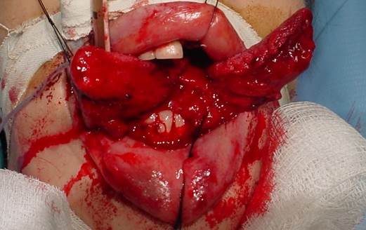
An adult patient is brought to the trauma bay after a high-speed motor-vehicle collision. There is major facial trauma, active oral bleeding, agitation, gurgling respirations, and inability to reliably protect the airway. Cervical-spine precautions are maintained. Continuous suction is required. After basic monitoring, suction, and preoxygenation have begun, a resuscitation-area clinical photograph is obtained during the airway examination. It shows the clinician-confirmed finding: the tongue is split in the midline nearly to its posterior aspect. Blood obscures the oropharynx, and associated facial fractures are suspected but not yet characterized.
 CASE 038
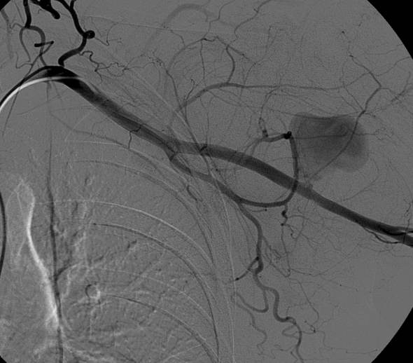
A 62-year-old patient falls approximately 4 meters from a roof, landing on the right shoulder and upper arm. On arrival, the patient is alert with a rapidly enlarging upper-arm hematoma and severe pain. Radial and ulnar pulses are palpable but diminished compared with the opposite side; the hand is warm with preserved motor and sensory function. A pressure dressing, analgesia, intravenous access, and appropriate resuscitation are initiated. After transient stabilization without refractory hypotension, CT angiography demonstrates suspected active arterial bleeding in the proximal arm. The patient is transferred promptly to interventional radiology for selective catheter angiography. The uploaded image is acquired during that angiographic procedure and is now available for interpretation.
 CASE 039
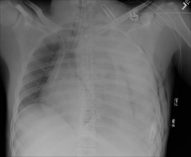
A previously healthy adult is brought directly to the trauma bay after a high-speed MVC. The patient is pale, confused, tachypneic, and has weak peripheral pulses. Initial blood pressure is 55/— mmHg and heart rate is 135/min. There is extensive left chest bruising and markedly diminished breath sounds on the left. The airway is secured with rapid-sequence intubation, large-bore access is obtained, and balanced blood-product resuscitation is started. A portable AP supine chest radiograph is obtained during ongoing resuscitation and is now available for review. No chest tube has yet been placed, and the patient remains severely unstable.
 CASE 040
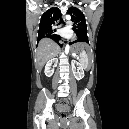
A 27-year-old woman is brought to the emergency department after an MVA. She was restrained and has left upper-quadrant tenderness without peritonitis. Initial airway and breathing assessments are reassuring. After analgesia and a small crystalloid bolus, her blood pressure and heart rate remain stable, and she is alert with no other immediately life-threatening injury identified. Because her physiology permits diagnostic imaging, a portal-venous-phase contrast CT of the chest, abdomen, and pelvis with multiplanar reconstructions is completed. The uploaded CT image is now available for interpretation.
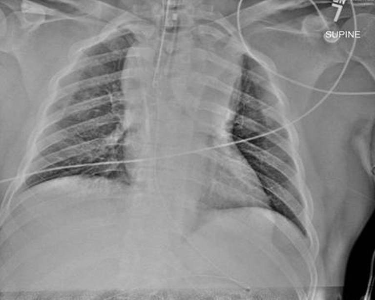 CASE 041
A 32-year-old patient is brought directly to the trauma bay after a high-speed motor-vehicle collision. Prehospital personnel report GCS 10 with agitation and inability to protect the airway, so rapid-sequence intubation was performed before transport. On arrival, the patient is mechanically ventilated, has palpable bilateral radial and femoral pulses, and has an adequate blood pressure after limited crystalloid. The team obtains a portable supine AP chest radiograph during the initial trauma survey. The image is available for review at this stage; the patient remains sufficiently perfused for monitored transport, with no uncontrolled external hemorrhage or other finding that mandates thoracotomy before CTA.
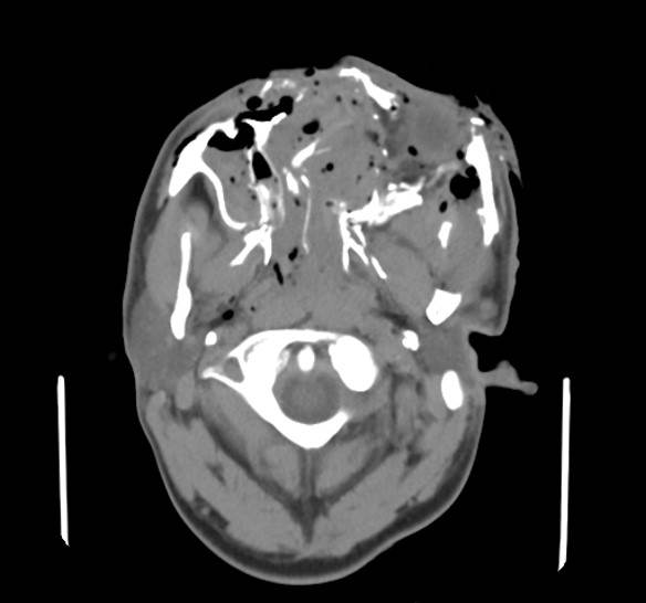 CASE 042
A 29-year-old patient is brought to a level I trauma center after a high-speed collision. The face struck the dashboard, and witnesses report brief loss of consciousness. On arrival, the patient is awake but agitated, with severe facial bleeding and inability to see from the left eye. Initial airway maneuvers and suction improve oxygenation, and blood pressure remains adequate after limited resuscitation. The patient is stable enough for imaging, so a CT of the head, face, and neck is obtained before operative management. The uploaded axial facial CT image is now available for interpretation. A cervical collar remains in place.
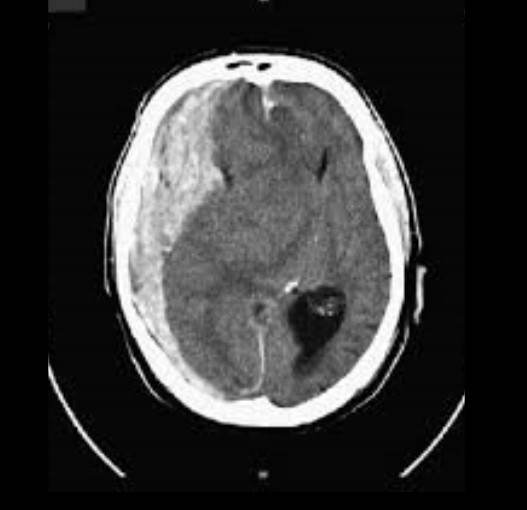 CASE 043
A 67-year-old man is brought to the emergency department after a high-speed collision. He was initially awake but became progressively somnolent during transport. On arrival, the airway is managed with cervical-spine precautions and assisted ventilation. After controlled initial resuscitation, blood pressure is 148/86 mmHg, heart rate 92/min, oxygen saturation 99% with assisted ventilation, and temperature 36.4°C. Neurologic examination shows GCS 7T, a right pupil measuring 5 mm and sluggishly reactive, a left pupil measuring 3 mm and reactive, and withdrawal only on the right. Family reports warfarin use for atrial fibrillation; initial INR is 2.4. He has no refractory hypotension or hypoxemia and is sufficiently stable for immediate diagnostic imaging.
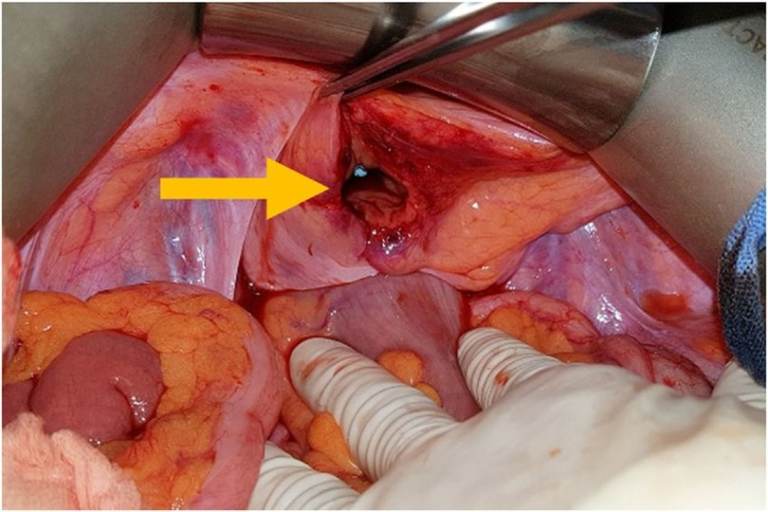 CASE 044
A front-seat passenger wearing a lap belt is brought after a frontal motor-vehicle collision. The patient has severe suprapubic and diffuse abdominal pain, microscopic hematuria, and guarding but remains hemodynamically stable after initial resuscitation. CT cystography demonstrates intraperitoneal contrast extravasation from the bladder dome, with free intraperitoneal fluid and no compelling evidence of an isolated extraperitoneal rupture. The patient remains stable after imaging and is taken to the operating room for planned abdominal exploration. The operative image becomes available after the abdominal field is exposed.
 CASE 045
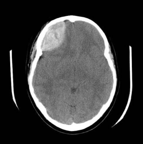
A 24-year-old previously healthy patient is brought to the emergency department after an unrestrained high-speed motor-vehicle collision. On arrival, the patient is awake but confused, with a right temporal scalp hematoma and one episode of emesis. Initial vital signs are stable, oxygenation is adequate, and there are no immediately threatening extracranial injuries. After cervical spine precautions, two large-bore IVs, analgesia, and neurologic reassessment, the patient remains sufficiently stable for urgent noncontrast head CT. The CT is obtained, and the uploaded image is now available for interpretation.
 CASE 046
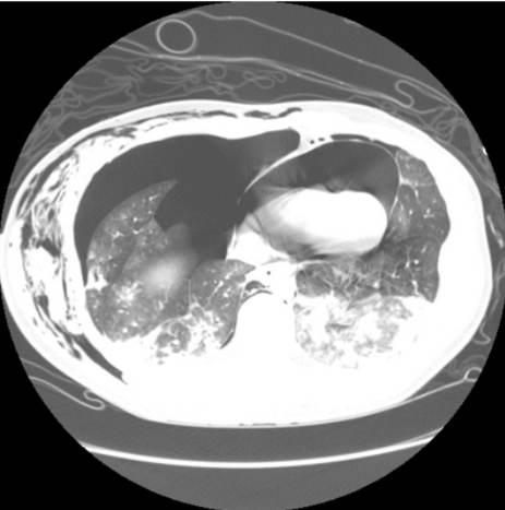
A 29-year-old patient is brought to the emergency department after a high-speed motor-vehicle collision. Initial airway and breathing assessment shows spontaneous respirations, severe right-sided chest pain, and diminished right breath sounds. After oxygen, analgesia, and cautious resuscitation, blood pressure is 108/72 mmHg, heart rate is 124/min, oxygen saturation is 91% on a nonrebreather mask, and mental status is intact. The patient has no refractory hypotension, severe altered mental status, or immediate peri-arrest physiology. After trauma-team assessment and initial stabilization, the patient is considered sufficiently stable for CT.
 CASE 047
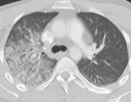
A 32-year-old patient is brought to the emergency department after a high-speed motor-vehicle collision. The patient was restrained and had a brief period of chest compression against the steering wheel. On arrival, the airway is patent, breath sounds are present bilaterally but reduced on the right, and there is right anterior chest tenderness with bruising. Initial oxygen saturation is 94% on a non-rebreather mask, blood pressure is 118/76 mmHg, and heart rate is 108/min. After analgesia, cervical protection, vascular access, and initial trauma evaluation, the patient remains sufficiently stable for CT. The chest CT has now been obtained and is available for review.
 CASE 048
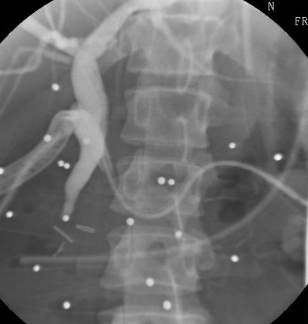
A 29-year-old man sustains a close-range shotgun blast to the upper abdomen. On arrival he is tachycardic and peritonitic, with multiple radiopaque pellets on plain radiography. He undergoes damage-control laparotomy with hemorrhage and contamination control, repair of associated hollow-viscus injuries, placement of drains, and temporary abdominal closure. The liver and porta hepatis are explored without an immediately recognized complete bile duct transection. He remains in the ICU with an open abdomen and a planned re-exploration. Over the next 72 hours, he develops progressive jaundice, dark urine, and increasing bilious output from a right upper-quadrant drain. He is resuscitated and remains sufficiently stable for further evaluation. Laboratory testing shows rising direct bilirubin and a cholestatic enzyme pattern. Ultrasound demonstrates intrahepatic and extrahepatic biliary dilation. After multidisciplinary review and appropriate anesthesia and surgical backup, ERCP is performed. The provided fluoroscopic image is obtained during cholangiography and is available to the learner at the first decision point.
 CASE 049
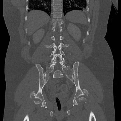
A 29-year-old restrained driver is brought to the trauma bay after a high-speed collision. The patient had transient hypotension in the field but responds to blood-product resuscitation. On arrival, the airway is patent, breathing is adequate, and pedal pulses are present bilaterally. The pelvis is painful, and the legs are shortened and held in abnormal rotation. After initial resuscitation, the patient remains sufficiently responsive and hemodynamically acceptable for CT. A contrast-enhanced trauma CT with multiplanar pelvic reconstructions is obtained. The displayed coronal CT image is now available for interpretation.
 CASE 050
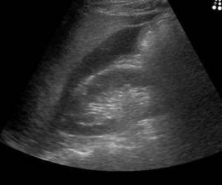
A motorcyclist is thrown from his bike and lands heavily on his right side. He arrives in the trauma bay alert, with an airway maintained, bilateral breath sounds, a palpable radial pulse, BP 115/74 mmHg, and HR 97/min. He reports right-upper-quadrant and right-flank pain. After initial trauma assessment and monitoring, an emergency physician performs a focused assessment with sonography for trauma (FAST). The uploaded image is the right-upper-quadrant FAST view and becomes available for review at this point. No other imaging has yet been obtained.
 CASE 051
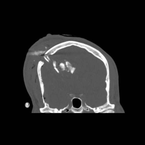
A 24-year-old patient is brought to the trauma bay after a witnessed close-range gunshot wound to the head. A pressure dressing is applied without obstructing the airway. On arrival, the patient is breathing spontaneously with oxygen saturation 96% on supplemental oxygen, blood pressure 132/78 mmHg, heart rate 104/min, and Glasgow Coma Scale score E2 V2 M5. The left pupil is 4 mm and sluggish; the right pupil is 3 mm and reactive. There is an obvious scalp defect with palpable calvarial disruption and blood at the wound. The patient is intubated for airway protection, cervical-spine precautions are maintained, and resuscitation produces stable oxygenation and blood pressure. Because the patient remains sufficiently stable for imaging, a noncontrast head CT and CT angiography are obtained in the emergency department. The uploaded coronal noncontrast CT is now available for review; the CTA remains pending formal interpretation.
 CASE 052
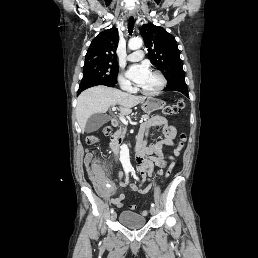
A restrained adult is brought to the emergency department after a high-speed motor-vehicle collision. There is a prominent lower-abdominal seat-belt sign and persistent right-sided abdominal pain. Initial airway and breathing assessments are normal. After balanced resuscitation, the patient remains sufficiently stable for imaging: heart rate 104/min, blood pressure 118/74 mmHg, warm extremities, and no peritonitis. A focused assessment does not show a large-volume free-fluid pattern. A portal-venous-phase contrast CT of the chest, abdomen, and pelvis is obtained before definitive treatment. The uploaded CT image is now available for review.
 CASE 053
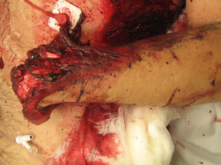
A 29-year-old motorcyclist is brought to the trauma bay after being dragged approximately 20 meters on asphalt. The patient was helmeted, has scattered abrasions, and is initially awake but distressed. A pressure dressing placed by first responders is saturated. The right hand is absent at the wrist or distal forearm, with a grossly contaminated mangled stump and extensive degloving. The amputated hand has not been located. There is no immediately obvious chest or abdominal injury. The trauma team performs a primary survey, applies direct pressure and a proximal tourniquet, obtains large-bore access, and begins warmed blood-product resuscitation. The patient has a palpable central pulse and transiently improves after hemorrhage control. A trauma-bay photograph obtained during this initial hemorrhage-control assessment is available for review.
 CASE 054
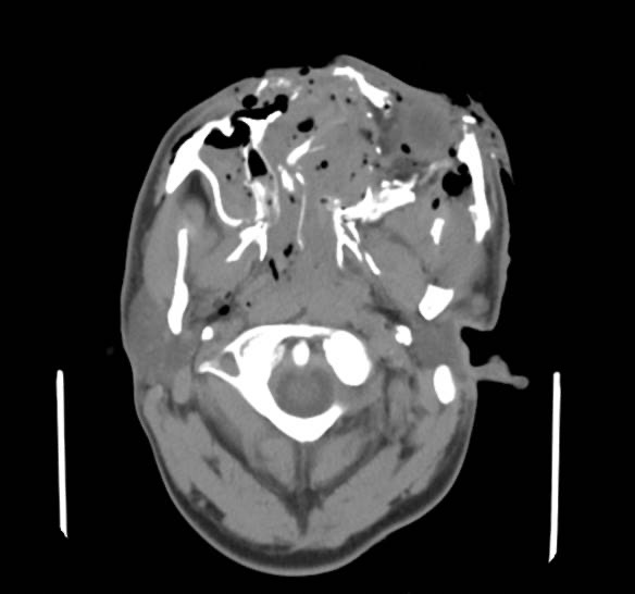
An adult patient is brought to the trauma bay after a high-speed motor-vehicle collision. There is profuse bleeding from complex facial lacerations and deformity of the midface. Direct pressure and hemostatic dressings have reduced but not stopped the bleeding. The patient is initially responsive but has gurgling respirations, blood in the oropharynx, and progressive facial swelling. A trauma airway team performs suction-assisted airway control with cervical-spine precautions, establishes large-bore access, begins balanced blood-product resuscitation, and obtains a contrast-enhanced CT of the head, face, and neck while the patient remains sufficiently responsive to resuscitation for transport. The displayed CT is now available for review.
 CASE 055
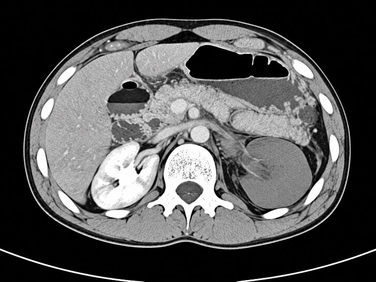
A previously healthy adult is brought after a high-speed motor-vehicle collision with ejection. Initial airway, breathing, circulation, and spine assessment is completed, and the patient receives blood products, warming, analgesia, and ongoing monitoring. The patient has epigastric and left-flank tenderness without peritonitis and remains sufficiently stable for contrast-enhanced CT during the early trauma evaluation. The CT is obtained before the supplied image is reviewed. The image is now available and shows the clinician-confirmed findings of traumatic pancreatic injury and a devascularized left kidney. The emphasis is deliberate preoperative planning rather than reflexive immediate operation.
 CASE 056
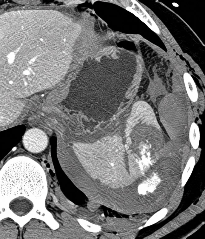
A 29-year-old restrained driver presents after a high-speed motor-vehicle collision with left upper quadrant pain and left shoulder discomfort. On arrival, airway and breathing are intact. Blood pressure is 122/76 mmHg, heart rate 96/min, and mental status is normal. After initial resuscitation with monitoring and limited crystalloid, vital signs remain stable. Focused abdominal examination shows left upper quadrant tenderness without peritonitis. Given the stable physiology, a contrast-enhanced CT of the abdomen and pelvis is obtained before the image is made available for review. The provided CT is now available for review.
 CASE 057
A 29-year-old patient arrives after a close-range stab wound to the left precordium. Prehospital personnel report transient loss of pulses with return of spontaneous circulation after a brief period of CPR. On arrival, the airway is secured, bilateral chest decompression has been performed for absent left breath sounds, and a massive transfusion protocol is initiated. The patient develops recurrent profound hypotension followed by pulselessness. The penetrating injury is in the left anterior chest, the collapse was witnessed shortly before arrival, and there are no signs of unsurvivable injury. The operating room is not immediately available, so the trauma team prepares for emergency resuscitative thoracotomy in the trauma bay. After temporary resuscitation and hemorrhage control, the patient will be transferred directly to the operating room with the chest open. The operative image becomes available after transfer and continued rib-spreader exposure.